Human Reproductive System
One Mark Questions
1. Name the cells that nourish the germ cells in the testes. Where are these cells located in the testes?
ANS:- ‑The cells that nourish the germ cells in the testes are called Sertoli cells. Sertoli cells are located in the germinal epithelium of the seminiferous tubules.
2. Write the function of the seminal vesicle.
ANS:- Seminal vesicle produces an alkaline secretion that helps to neutralize the acidic environment of the male urethra as well as that of the female reproductive tract.
3. Write the location and function of Sertoli cells in humans.
ANS:- Refer to answer 1.
4. What is the function of Leydig’s cells?
ANS:- Leydig’s cells synthesize and secrete testicular hormones called androgens.
5. When do the oogenesis and the spermatogenesis initiate in human females and males respectively?
ANS:- Oogenesis begins during the embryonic development stage while spermatogenesis begins during puberty.
6. Write the function of the acrosome of human sperm.
ANS:- Acrosome contains enzymes called spermlysins that are used to contact and penetrate the ovum at the time of fertilization.
7. List the changes the primary oocyte undergoes in the tertiary follicular stage in the human ovary.
ANS:- The primary oocyte grows in size and completes its first meiotic division within the tertiary follicle.
8. Sperms have a tail whereas eggs do not. Why so?
ANS:- Sperms are tailed whereas eggs do not as sperms have to move (tail helps in locomotion) through the cervix, uterus, and Fallopian tube to reach the egg already present there.
9. Write the physiological reason, why a woman generally cannot conceive a child after 50 years of age?
ANS:- A woman generally cannot conceive a child after 50 years of age as at this age menopause occurs. It is a phase in a woman’s life when ovulation and menstruation stops.
10. Mention the function of zona pellucida.
ANS:- During fertilization, a sperm comes in contact with the zona pellucida layer of the ovum which induces changes in the membrane that block the entry of additional sperms. So, zona pellucida ensures that only one sperm can fertilize an ovum.
11. How does the sperm penetrate through the zona pellucida in the human ovum?
ANS:- The sperm penetrate through the zona pellucida by releasing sperm lysins.
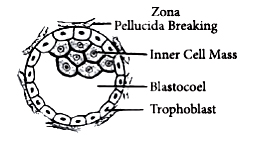
12. Mention the function of the trophoblast in human embryos.
ANS:- Trophoblast helps to provide nutrition to the embryo.
13. Name the embryonic stage that gets implanted in the uterine wall of a human female.
ANS:- Blastocyst.
14. Write the function of oxytocin.
ANS:- Oxytocin acts on the uterine muscle causing contractions that help in the expulsion of the baby out of the uterus through the birth canal.
15. What stimulates the pituitary to release the hormone responsible for parturition? Name the hormone?
ANS:- ‑The signals from the fully developed foetus and placenta trigger the release of oxytocin from the maternal pituitary. Oxytocin is responsible for parturition.
16. Mention the difference between spermatogenesis and spermiation.
ANS:- The process of formation of sperms is called spermatogenesis while the release of sperms from the seminiferous tubules is called spermiation.
17. Where is acrosome present in humans? Write its function.
ANS:- Acrosome present in the head of sperm. It helps in dissolving the layer of the egg during fertilization.
18. When does oogenesis begin?
ANS:- Acrosome is present in the head of the human sperm.
19. How is the entry of only one sperm and not many ensured into an ovum during fertilization in humans?
ANS:- Depolarisation of the egg plasma membrane by binding of sperm to it checks additional sperms from entering into it.
20. Explain the function of the umbilical cord.
ANS:- The placenta is connected to the embryo through an umbilical cord which helps in the transport of substances to and from the embryo.
Two Mark Questions
1. Why are the human testes located outside the abdominal cavity? Name the pouch in which they are present.
ANS:- The human testes are located outside the abdominal cavity within a pouch called the scrotum. The scrotum helps in maintaining the low temperature of the testes (2- 2.5°C) lower than the normal internal body temperature, necessary for spermatogenesis.
2. Write the location and functions of the following in human testes :
(a)Sertoli cells
(b) Leydig’s cells.
ANS: - (a) The cells that nourish the germ cells in the testes are called Sertoli cells. Sertoli cells are located in the germinal epithelium of the seminiferous tubules.
(b) Leydig’s cells synthesize and secrete testicular hormones called androgens.
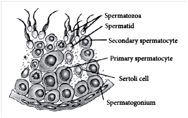
3. Draw a labeled diagram of the sectional view of the seminiferous tubule of a human.
ANS:- Sectional view of the human seminiferous tubule is as follows:
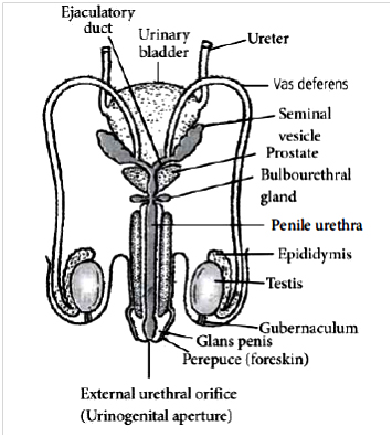
4. The diagram below shows the human male reproductive system (one side only).
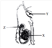
(a) Identify ‘X’ and write its location in the body.
(b) Name the accessory gland ‘Y’ and its secretion.
(c) Name and state the function of ‘Z’
ANS:- (a) X is testis. It is located outside the abdominal cavity within a pouch called the scrotum.
(b) Y is a seminal vesicle. It produces an alkaline secretion that forms 60% of the volume of the semen.
(c) Z is the epididymis. It stores the sperms and also secretes a uid which nourishes the sperms.
5. Draw a labeled diagram of the human female reproductive system.
ANS:-
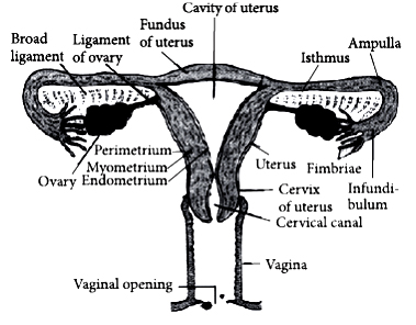
6. Explain the functions of myometrium and endometrium in human females.
ANS:- Myometrium is the middle thick layer of smooth muscles fibers that bring about contraction of the uterus during the delivery of the baby. The endometrium is the inner glandular layer that undergoes cyclical changes during the menstrual cycle.
7. Mention the names and the characteristics of different uterine wall layers in humans. Which one of them undergoes cyclic changes during menstrual cycles?
ANS:- The wall of the uterus is composed of three layers of tissues. The perimetrium is an outer thin covering of the peritoneum. The myometrium is a middle thick layer of smooth muscle fibers that shows strong contraction during delivery of the baby. The endometrium is the inner glandular layer that lines the uterine cavity. The endometrium undergoes cyclical changes during the menstrual cycle.
8. Write two major functions each of the testis and ovary.
ANS:- Functions of the testis:
(i) It produces sperms (male gametes).
(ii) It secretes male sex hormones i.e., androgens(e.g., testosterone).
Functions of the ovary :
(i) It produces ova (female gametes).
(ii) It secretes female sex hormones e.g., estrogen and progesterone.
9. Explain the hormonal regulation of the process of spermatogenesis in humans.
ANS:- During spermatogenesis, gonadotropin-releasing hormone (GnRH) is secreted by the hypothalamus, which stimulates the anterior pituitary gland to secrete luteinizing hormone (LH) and follicle-stimulating hormone (FSH). LH acts on the Leydig’s cells of the testes to secrete testosterone while FSH acts on Sertoli cells of the seminiferous tubules of the testes to secrete androgen binding protein (ABP) and inhibin. ABP concentrates testosterone and inhibin suppresses FSH synthesis. FSH also acts on spermatogonia to stimulate sperm production.
10. Draw a detailed structure of human sperm. Label the cellular components.
ANS:- 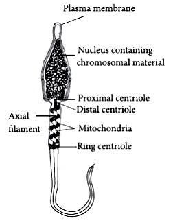
11. Identify A, B, C, and D with reference to gametogenesis in humans, in the ow chart given below.
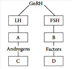
ANS:- A – Leydig’s cells, B – Sertoli cells, C–Spermatogenesis, D – Spermiogenesis.
12. Name the labels a, b, c, d, e, f in the diagram of the seminiferous tubule.
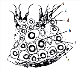
ANS:- In the given diagram of seminiferous tubule:-
a–Spermatogonium, b–Primary spermatocyte, c–Spermatid, d–Secondary spermatocyte,
e–Spermatozoa, f–Sertoli cell
13. Differentiate between menarche and menopause.
ANS:- Beginning of menstruation or first menstruation is called menarche that occurs at puberty. When the menstrual cycle ceases around the age of 50 years, it is called menopause.
14. Name the hormones influencing
(a) ovulation,
(b) development of corpus luteum..
ANS:- (a) On the 14th day of the menstrual cycle,s urge in LH causes rupturing of Graafian follicle and release of an ovum (ovulation).
(b) After ovulation, the remaining cells of the ovarian follicles are stimulated by LH to develop the corpus luteum.
15. Where does fertilization occur in humans? Explain the events that occur during this process.
ANS:- The motile sperms swim rapidly, pass through the cervix, enter into the uterus and finally reach the junction of the isthmus and ampulla (ampullaryisthmic junction) of the Fallopian tube. The ovum released by the ovary is also transported to the ampullary-isthmic junction where fertilization takes place. During fertilization, a sperm comes in contact with the zona pellucida layer of the ovum and induces changes in the membrane that block the entry of additional sperms. ‑The secretions of the acrosome help the sperm enter into the cytoplasm of the ovum through the zona pellucida and the plasma membrane. This induces the completion of the meiotic division of the secondary oocyte. The second meiotic division is also unequal and results in the formation of a second polar body and haploid ovum (ootid). Soon the haploid nucleus of the sperms and that of the ovum fuse together to form a diploid zygote.
16. Name the embryonic stage that gets implanted in the human female. Explain the events that occur during this process.
ANS:- Blastocyst gets implanted in the human females. In a blastocyst, the blastomeres are arranged into an outer layer called trophoblast and an inner group of cells called the inner cell mass. The trophoblast then gets attached to the endometrium and the inner cell mass gets dierentiated as the embryo. After attachment, the uterine cells divide rapidly and cover the blastocyst. As a result, the blastocyst becomes embedded in the endometrium of the uterus. This whole phenomenon is called implantation and it leads to pregnancy.
17. Why is parturition called a neuroendocrine mechanism?
ANS:- Process of parturition is induced by both the neural system and endocrine system therefore, it is called a neuroendocrine mechanism. The signals for parturition originate from the fully developed foetus and the placenta which induce mild uterine contractions called foetal ejection reflex. This triggers the release of oxytocin from the maternal pituitary. Oxytocin acts on the uterine muscle and causes stronger uterine contractions that lead to the expulsion of the baby out of the uterus through the birth canal.
18. How is milk production regulated by hormones in human females? Explain.
ANS:- Secretion and storage of milk generally begin after birth of a young one, usually within 24 hours under the influence of the hormone prolactin (PRL) secreted by the anterior lobe of the pituitary gland. However, the ejection of milk is stimulated by the hormone oxytocin (OT) released from the posterior lobe of the pituitary gland.
19. Comment on the role of the placenta as an endocrine gland.
ANS:- The placenta acts as an endocrine gland and secretes the following hormones :
(i) Human chorionic gonadotropin (hCG)
(ii) Human chorionic somatomammotropin (hCS)
(iii) Progesterone
(iv) Estrogen
(v) Relaxin
(vi) Chorionic thyrotropin and
(vii) Chorionic corticotropin
The hCG stimulates and maintains the corpus luteum to secrete progesterone until the end of pregnancy. The hCS stimulates the growth of the mammary glands during pregnancy. Relaxin facilitates parturition (the act of birth) by softening the connective tissues of the pubic symphysis. The level of hormones like estrogen, progesterone, etc. is increased in maternal blood during pregnancy. Increased production of these hormones is necessary for supporting the fetal growth, metabolic changes in mother, and maintenance of pregnancy.
20. When and where do chorionic villi appear in humans? State their function. How is milk production regulated by hormones in human females? Explain.
ANS:- (a)‑The signals for parturition originate from the fully developed foetus and the placenta which induce mild uterine contractions called foetal ejection reflex.
(b) After birth, the breast first releases milk is called colostrum for 2 or 3 days. This is a thin, yellowish, fluid, often called foremilk. It contains cells from the alveoli and is rich in protein (lactalbumin & lactoprotein), antibodies, but low in fat. Breast-feeding during the initial period of infant growth is recommended by doctors for bringing up a healthy baby.
Three Mark Questions
1. Name and explain the role of the inner and middle walls of the human uterus.
ANS:- The inner glandular wall of the uterus is known as the endometrium.
Role - During the menstrual cycle, the endometrium wall grows into a thick, vascular (blood vessel-rich) glandular layer. This condition of the endometrium favors the implantation of the foetus. If fertilization does not occur, the endometrium is shed during the hemorrhagic phase of the menstrual cycle. The middle wall of the uterus is known as the myometrium.
Role - It consists of smooth muscles. It exhibits contraction during the delivery of the baby.
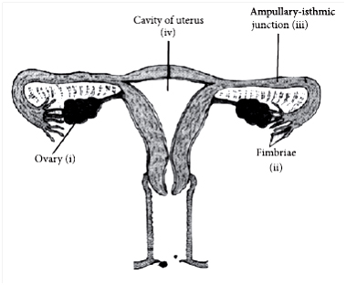
2. Draw a diagrammatic sectional view of the female reproductive system of humans and label the parts
(i) where the secondary oocytes develop
(ii) which helps in the collection of ovum after ovulation
(iii) where fertilization occurs
(iv) where implantation of the embryo occurs.
ANS:- Diagrammatic sectional view of the female reproductive system is as follows:
.
3. Explain the hormonal control of spermatogenesis in humans.
ANS:- During spermatogenesis, gonadotropin-releasing hormone (GnRH) is secreted by the hypothalamus, which stimulates the anterior pituitary gland to secrete luteinizing hormone (LH) and follicle-stimulating hormone (FSH). LH acts on the Leydig’s cells of the testes to secrete testosterone while FSH acts on Sertoli cells of the seminiferous tubules of the testes to secrete androgen binding protein (ABP) and inhibin. ABP concentrates testosterone and inhibin suppresses FSH synthesis. FSH also acts on spermatogonia to stimulate sperm production.
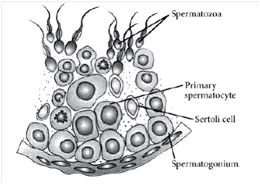
4. Draw a diagrammatic sectional view of a human seminiferous tubule and label Sertoli cell, primary spermatocyte, spermatogonium, and spermatozoa in it.
ANS:- Sectional view of the human seminiferous tubule is as follows:
5. Draw a diagrammatic sectional view of the human ovary to show the development of follicles and ovulation. Label the different stages in the diagram.
ANS:- Diagrammatic sectional view of human ovary showing the development of follicles and ovulation is as follows:
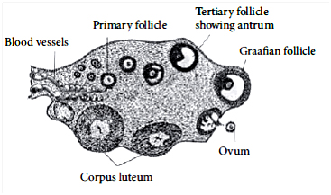
6. Draw a sectional view of the human ovary. Label the following parts :
(i) Primary follicle
(ii) Ovum
(iii) Graafian follicle
(iv) Corpus luteum.
ANS:- Refer to answer 5.
7. Explain the events in a normal woman during her menstrual cycle on the following days:
(a) Ovarian event from 13-15 days
(b) Ovarian hormones level from 16 to 23 days
(c) Uterine events from 24 to 29 days.
ANS:- (a) During 13-15 days, FSH stimulates the ovarian follicle to secrete estrogens that further stimulate the proliferation of the endometrium of the uterine wall. On the 14th day, LH surge causes ovulation.
(b) From 16 to 23 days, the corpus luteum secretes progesterone which is required for the maintenance of endometrium. In the absence of fertilization, the corpus luteum degenerates causing disintegration of the endometrium leading to menstruation that takes 3-5 days.
(c) During 24 to 29 days (luteal phase of 15 to 28 days), the luteinizing hormone (LH) is secreted by the anterior lobe of the pituitary gland. LH causes ovulation. The remaining cell of the ovarian follicles is stimulated by the LH to develop corpus luteum. The corpus luteum secretes a large amount of progesterone. Progesterone stimulates the uterine glands to produce an increased amount of watery mucus. During the secretory phase, there is also a similar increase in the secretion of watery mucus by the vaginal glands and by the glands of the Fallopian tubes. Progesterone is also essential for the maintenance of the endometrium which is necessary for implantation of the fertilized ovum and other events of pregnancy. In the absence of fertilization, the corpus luteum degenerates. This causes disintegration of the endometrium leading to menstruation marking a new cycle.
8. Explain the events in a normal woman during her menstrual cycle on the following days :
(a) Pituitary hormone levels from 8 to 12 days.
(b) Uterine events from 13 to 15 days.
(c) Ovarian events from 16 to 23 days.
ANS:- (a) From 8-12 days (follicular phase), the level of gonadotropins (LH and FSH) increase gradually and stimulate follicular development as well as secretion of estrogens by growing follicles.
(b) From 13 to 15 days, the endometrium becomes thicker by rapid cell multiplication and there is an increase in uterine glands and blood vessels.
(c) On the 14th day of the menstrual cycle, the surge in LH causes rupturing of Graafian follicle and release of an ovum (ovulation).
After ovulation, the remaining cells of the ovarian follicles are stimulated by LH to develop the corpus luteum.
9. When does the corpus luteum degenerate?
Explain the immediate consequences of its degeneration in human females.
ANS:- In the absence of fertilization, the corpus luteum degenerates. Degeneration of the corpus luteum leads to a decrease in the production of progesterone. As progesterone is needed for the maintenance of the endometrium, its reduction leads to the disintegration of the endometrium thus causing menstruation.
10. Briefly explain the events of fertilization and implantation in an adult human female.
ANS:- The events of fertilization in human females are:
(i) Acrosomal reaction: After ovulation, the secondary oocyte reaches the Fallopian tube. The capacitated sperm releases hydrolytic enzymes (sperm lysins) present in the acrosome when it comes in contact with the surface of the egg covering. Important sperm lysins are (i) hyaluronidase that acts on the ground substances of follicle cells, (ii) corona penetrating enzyme that dissolves corona radiata, and (iii) zona lysine or acrosin that helps to digest the zona pellucida. Due to acrosomal reaction, the plasma membrane of sperm fuses with that of the secondary oocyte, and depolarisation of the oocyte membrane occurs.
(ii) Cortical reaction: Immediately after the fusion of sperm and egg plasma membranes, the egg shows a cortical reaction to further check the entry of more sperms. In this reaction, the cortical granules present beneath the ovum’s plasma membrane fuse with the same and release their contents (enzymes) between it and zona pellucida. These enzymes harden the zona pellucida, which now functions as the sure block to polyspermy.
(iii) Sperm entry: The egg extends around the entering sperm, finger-like processes, called microvilli, which constitute a fertilization cone. The latter takes the entire sperm into the egg. The distal centriole of the sperm divides and forms two centrioles to generate the mitotic spindle for cell division.
(iv) Karyogamy: The sperm entry stimulates the egg (secondary oocyte) to resume and complete the suspended meiosis - II. This produces a haploid mature ovum and a second polar body. ‑The head of sperm separates from the middle piece and tail to become a male pronucleus and the nucleus of the ovum is called the female pronucleus. The second polar body and sperm tail degenerate. Mixing up the chromosomes of a spermatozoon and an ovum is called karyogamy or amphimixis. This completes the act of fertilization. The fertilized ovum is now a diploid cell having 23 pairs of chromosomes and is termed a zygote. The events of implantation are discussed as follows:
Implantation is the attachment of the blastocyst to the uterine wall. It occurs after 7 days of fertilization. As the zygote moves towards the uterus, it undergoes series of mitotic divisions known as cleavage and forms 2,4,8,16 daughter cells called blastomeres. The embryo with 8 blastomeres is called a morula. The morula transforms into a blastocyst. In a blastocyst, the blastomeres are arranged into an outer layer called trophoblast and an inner group of cells called the inner cell mass. The trophoblast then gets attached to the endometrium and the inner cell mass gets dierentiated as the embryo. After attachment, the uterine cells divide rapidly and cover the blastocyst. As a result, the blastocyst becomes embedded in the endometrium of the uterus. This whole phenomenon is called implantation and it leads to pregnancy.
11. List and explain the events that follow the journey of the human zygote until its implantation.
ANS:- Refer to answer 10.
12. (a) How is the placenta formed in the human female?
(b) Name any two hormones which are secreted by it and are also present in a nonpregnant woman.
ANS:- (a) After implantation, finger-like projections appear on the trophoblast called chorionic villi which are surrounded by the uterine tissue and maternal blood. The chorionic villi and uterine tissue become interdigitated with each other and jointly form a structural and functional unit between the developing embryo (foetus) and maternal body called the placenta, which facilitates the supply of oxygen and nutrients to the embryo and also removal of carbon dioxide and excretory waste materials produced by the embryo. Placenta also acts as an endocrine tissue and produces several hormones essential for supporting the foetal growth, metabolic changes in the mother, and maintenance of pregnancy.
(b)Estrogen and progesterone.
13. Describe the process of parturition in humans.
ANS:- The signals for parturition originate from the fully developed foetus and the placenta which induce mild uterine contractions called foetal ejection reex. After birth, the breast's first release of milk is called colostrum for 2 or 3 days. This is a thin, yellowish, fluid, often called foremilk. It contains cells from the alveoli and is rich in protein (lactalbumin & lactoprotein), antibodies, but low in fat. Breast-feeding during the initial period of infant growth is recommended by doctors for bringing up a healthy baby.
Five Mark Questions
1. (a) Draw a labeled diagrammatic view of the human male reproductive system.
(b) Differentiate between :
(i) Vas deferens and vasa Defferentia.
(ii) Spermatogenesis and spermiogenesis.
ANS:-(a) Diagrammatic view of the human male reproductive system is as follows.
(b) (i) Differences between vasa efferentia and vasa deferentia are as follows:
|
|
Vasa efferentia |
Vasa deferentia |
|
01 |
They arise from the rete testis. |
They arise from the cauda epididymis. |
|
02 |
They vary from 15 to 20 in number. |
They are only 2 in number. |
|
03 |
Vasa efferentia are fine. |
Vasa deferentia are thick. |
|
04 |
Their lining bears many ciliated cells. |
Their lining has many stereocilia. |
|
05 |
It carries spermatozoa from the rete testis to the epididymis. |
It carries spermatozoa from the cauda epididymis to the ejaculatory duct. |
(ii) Differences between spermatogenesis and spermiogenesis are as follows:
|
|
Spermatogenesis |
Spermiogenesis |
|
01 |
It is the process of formation of haploid spermatozoa from germinal cells. |
It is the process of dierentiation of spermatozoon from a spermatid. |
|
02 |
It involves the conversion of a diploid structure into haploid structures. |
It changes a haploid structure into another haploid structure. |
|
03 |
There is growth and division during spermatogenesis. |
There is reconstruction during spermiogenesis. Divisions and growth are absent. |
|
04 |
No organelle is lost. |
Golgi bodies are lost during spermiogenesis. |
|
05 |
A spermatogonium forms four spermatozoa. |
Here a spermatid forms a single spermatozoon. |
|
06 |
It consists of multiplication phase, spermatocytogenesis, maturation phase, and dierentiation phase |
It consists of only dierentiation phase. |
2. Explain the development of a secondary oocyte (ovum) in a human female from the embryonic stage up to its ovulation. Name the hormones involved in this process.
ANS:- The process of formation of a mature female gamete (ovum) is called oogenesis. It occurs in the ovaries. It consists of three phases: multiplication, growth, and maturation.
(i) Multiplication phase: In the foetal development, certain cells in the germinal epithelium of the ovary of the foetus are larger than others. These cells divide by mitosis, producing a couple of million egg mother cells or oogonia in each ovary of the foetus. The oogonia multiply by mitotic divisions forming the primary oocytes.
(ii) Growth phase: This phase of the primary oocyte is very long. The oogonium grows into a large primary oocyte by taking food from the surrounding follicle cells.
(iii) Maturation phase: Each primary oocyte undergoes two maturation divisions, the first meiotic and the second meiotic. In the first, meiotic division, the primary oocyte divides into two very unequal haploid daughter cells a large secondary oocyte and a very small first polar body or polocyte. In the second maturation division, the first polar body may divide to form two second polar bodies. The secondary oocyte again divides into unequal daughter cells, a large ootid and a very small second polar body. The ootid grows into a functional haploid ovum. Thus from one oogonium, one ovum and three polar bodies are formed. The polar bodies take no part in reproduction and, hence, soon degenerate. In humans, the ovum is released from the ovary in the secondary oocyte stage, this process is called ovulation. Estrogen & Progesterone.
3. Describe the events of spermatogenesis with the help of a schematic representation.
ANS:- Spermatogenesis is the process of formation of haploid spermatozoa from diploid spermatogonia inside the testes of the male. It includes the following three phases :
(i) Multiplication phase – At sexual maturity, the undierentiated primordial germ cells divide several times by mitosis to produce a large number of spermatogonia or sperm mother cells. Spermatogonia (2N) are of two types : type A spermatogonia and type B spermatogonia. Type A spermatogonia serve as the stem cells which divide to form the second type of spermatogonia whenever required. Type B spermatogonia are progenitor cells that function as precursors of spermatozoa.
(ii) Growth phase - Each type B spermatogonium actively grows to a larger primary spermatocyte by obtaining nourishment from the nursing cells.
(iii) Maturation phase - Each primary spermatocyte undergoes two successive divisions, called maturation divisions. The first maturation division is reductional or meiotic. Hence, the primary spermatocyte divides into two haploid daughter cells called secondary spermatocytes. Both secondary spermatocytes now undergo second maturation division which is an ordinary mitotic division to form four haploid spermatids, by each primary spermatocyte.
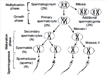
4. Give a schematic representation of spermatogenesis in humans.
ANS:- Refer to answer 3.
(b) At which stage of life does gametogenesis begin in humans males and females respectively?
ANS:- Oogenesis begins during the embryonic development stage while spermatogenesis begins during puberty.
5. (a) Explain the menstrual phase in a human female. State the levels of ovarian and pituitary hormones during this phase.
(b) Why is the follicular phase in the menstrual cycle also referred to as the proliferative phase? Explain.
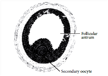
(c) Explain the events that occur in a Graafian follicle at the time of ovulation and thereafter.
(d) Draw a Graafian follicle and label antrum and secondary oocyte.
ANS:- (a) In a 28-day menstrual cycle, the menses takes place on days 3-5. The production of LH from the anterior lobe of the pituitary gland is considerably reduced. The withdrawal of this hormone causes degeneration of the corpus luteum and, therefore, progesterone production from the ovary is reduced. Production of estrogens from the ovary is also reduced in this phase. The endometrium of the uterus breaks down and menstruation begins. The cells of endometrium secretions, blood, and the unfertilized ovum constitute the menstrual flow.
(b) During the follicular phase, follicle-stimulating hormone (FSH) stimulates the ovarian follicle to secrete estrogens, which in turn stimulate the proliferation of the endometrium of the uterine wall. As a result, the endometrium becomes thicker by rapid cell multiplication and is accompanied by an increase of uterine gland and blood vessels. Hence, this phase is also referred to as a proliferative phase.
(c) At the time of ovulation, rapid secretion of LH induces rupturing of Graafian follicle, thereby releasing ovum. After ovulation has taken place, LH stimulates cells of the ovarian follicle to develop corpus luteum. Corpus luteum secretes large amount of progesterone.
(d)The structure of a mature Graafian follicle is as follows:
6. Study the graph given below and answer the questions that follow :
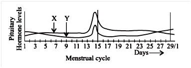
(a) Name the hormones ‘X’ and ‘Y’.
(b) Identify the ovarian phases during a menstrual cycle
(i) 5th day to 12th day of the cycle
(ii) 14th day of the cycle
(iii) 16th day to 25th day of the cycle
(c) Explain the ovarian events (i), (ii), and (iii) under the influence of hormones ‘X’ and ‘Y’.
ANS:- (a) X – Luteinising hormone (LH) Y – Follicle-stimulating hormone (FSH)
(i) Proliferative phase
(ii) Ovulatory phase
(iii) Luteal phase
(c) (i) During the follicular phase, FSH stimulates the ovarian follicle to secrete estrogen. Estrogen stimulates the proliferation of the endometrium of the uterine wall.
(ii) During the ovulatory phase, both LH and FSH attain a peak level. Rapid secretion of LH induces rupturing of Graafian follicle and thereby the release of the ovum.
(iii) During the luteal phase, LH causes the remaining cells of the ovarian follicles to develop corpus luteum.
7. Mention the site of fertilization of a human ovum. List the events that follow in sequence until the implantation of the blastocyst.
ANS:- Fertilisation occurs in the ampullary-isthmic junction of the Fallopian tube. Refer to answer 4.
8. List and explain the events that follow the journey of the human zygote until its implantation.
ANS:- Refer to answer 3.
9. (a) Explain the process of fertilization of an ovum in humans. Trace the events that occur after fertilization up to the implantation of the blastocyst.
ANS:- ANS:- Refer to answer 3.
(b) Draw a labeled diagram of a human blastocyst.
-------------------Compiled by:- AKB Sir-------------

